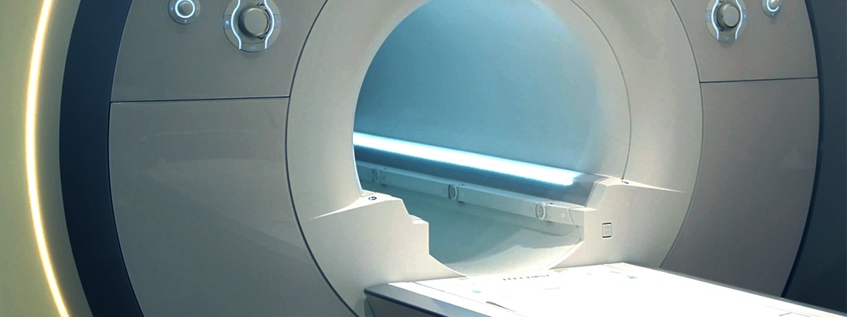
Accurate imaging is often the first step to healing. This is true not least in cancer medicine. While the parent tumor is usually easy to see, metastases can be as small as the head of a pin. To ensure successful therapy, however, we also need to find the tiniest metastases. This is achieved with positron emission tomography. It produces its images using a small amount of radiating particles that are administered to the patient mixed with glucose.
Greedy cells
Tumor cells need a lot of energy and greedily consume the sugar substance. With this, the radioactive particles accumulate in the metastases. The spatially concentrated radiation is then clearly visible in the PE tomogram.This type of diagnostics uses comparatively harmless, only weakly radiating substances such as the fluorine isotope 18F. Its half-life is just 110 minutes, meaning it has lost practically all its radioactivity after just one day. This means it has to be freshly produced just before it is used in a particle accelerator, the cyclotron.
Pinpoint proton bombardment
Inside the cyclotron is a vacuum chamber. There, negatively charged hydrogen ions are accelerated along a spiral path by means of strong electric fields. Once they reach the end, they fly through a thin graphite foil, where they lose their electrons and become positively charged protons. The reversal of the charge also directs it on a straight path.The resulting proton beam hits the material, triggers a nuclear reaction in it and produces the required isotopes. The vacuum is necessary so that neither the negative ions nor the positive protons are deflected by interfering particles on their way. The BUSCH Group offers suitable solutions for vacuum generation. Without a vacuum, the ions and protons would be deflected from their predetermined path and lose their energy. In addition, fluorine, which is extremely reactive, must not come into contact with other elements such as atmospheric oxygen.
